Laparoscopic radical left nephrectomy

Laparoscopic radical nephrectomy, also known as a kidney removal – is a standard accepted way to treat kidney cancer I the 2nd stage or further advanced, and sometimes even in the 1st stage.
This video has minimal amount of editing and shows all stages of the procedure.
Laparoscopic left radical nephrectomy
Description of surgical procedure:
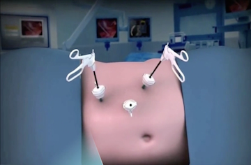
- We use 4 trocars using a standard diamond shaped pattern. NB! Very often during the incorrect positioning of the left trocar can present a problem. Our opinion is that it should be positioned along the pararectal line as high as possible which will ensure that the angle between the instrument and the renal pedicle will be close to 45 degrees which will allow for the most comfortable handling of the instrument.
- Using ultrasound scissors the descending colon is mobilised along the Toldt's fascia. We think it is important to gauge the non-vascular gap between the Toldt's fascia and gerota's fascia. Otherwise there is a risk of damaging blood vessels of the mesenterium of the descending colon or damage to the gerota’s fascia which would interfere with the principle of minimum intervention. I prefer to use a laparoscopic grasper in order to stop capillary haemorrhage and consider this instrument most useful for both coagulation and delicate dissection and traction.
- Following mobilisation of the descending colon it is essential to locate the ureter and the gonadal vein. The latter two are the points of reference for locating the renal helum.
- The next stage consists of isolating the elements of the renal helum – the artery and the vein. Both need to be clipped after mobilising. Clamping the blood vessels surrounded by fatty tissue is a mistake as it is very hard to maintain control over them. We prefer to use a soft tourniquet to gently manipulate the kidney vein.
- After isolating and clipping the kidney artery and kidney vein the ureter needs to also be isolated and clamped. We use Hem-O-Lok clamps for use on both the kidney vessels and vein.
- The isolation of the kidney is carried out on the rear surface between the rear folium of the gerota's fascia and the psoas. Gradual isolation of the kidney follows.
- The kidney and the surrounding tissue are placed inside an endobag.
- A draining tube is then inserted into the retroperitoneum.
- Lately we prefer to restore the abdominal membrane along the line of the Toldt's fascia because we consider this manipulation to be less likely to cause abdominal adhesions and aids faster restoration of gut mobility.
- The agent placed inside the endobag is removed via a small incision 4-5cm below the navel or alternatively, if the patient had had an appendectomy previously it can be removed via the appendectomy incision.

